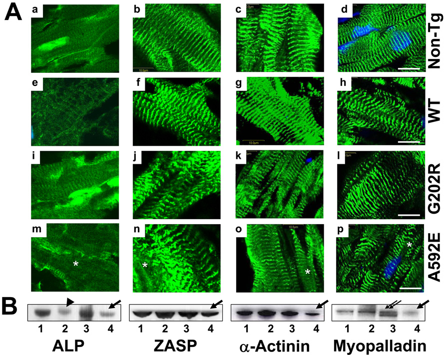
Figure 7. Comparative analysis of Z-disk proteins in mouse hearts.
Panel A. Representative cardiac sections stained for ALP (a, e, i and m), ZASP (b, f, j and n), α-actinin-2 (c, g, k and o), and myopalladin (d, h, l and p) from a non-Tg (a–d), WT (e–h), G202R (i–l) and A592E (m–p) mouse. Proteins are green and nuclei are blue (DAPI). Bar=10µm. Panel B. Western blotting of Z-disk proteins in non-Tg (1), WT (2), G202R (3) and A592E (4) hearts. Beta-Actin is used as a loading control (Figure 5B). Arrows and arrowhead indicate downregulation, asterisk indicates cleavage.

