Fig. 5.
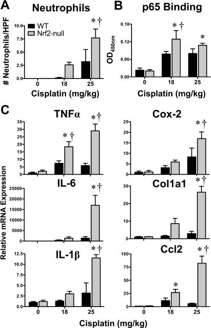
Neutrophil infiltration, p65 NFκB binding, and inflammatory mediator mRNA expression in kidneys of wild-type and Nrf2-null mice after cisplatin treatment. A, the number of neutrophils in three nonoverlapping high-powered fields were quantified in hematoxylin and eosin-stained kidney sections from vehicle or cisplatin (18 or 25 mg/kg)-treated wild-type and Nrf2-null mice on day 4. B, binding of kidney nuclear extracts from vehicle and cisplatin (18 or 25 mg/kg)-treated mice to p65 NFκB DNA-response element using an ELISA-based format. Data are presented as optical density (OD) at 450 nm. C, messenger RNA expression of tumor necrosis factor α, IL-6, IL-1β, cyclooxygenase 2, Col1a1, and Ccl2 was quantified using total kidney RNA from control and cisplatin (18 or 25 mg/kg)-treated wild-type and Nrf2-null mice on day 4. Data (n = 3–9) are presented as means ± S.E. Messenger RNA data were normalized to wild-type control mice. Black bars represent wild-type mice, and gray bars represent Nrf2-null mice. * represents statistically significant differences (p < 0.05) compared with genotype control mice. † represents a statistically significant difference (p < 0.05) from cisplatin-treated wild-type mice.

