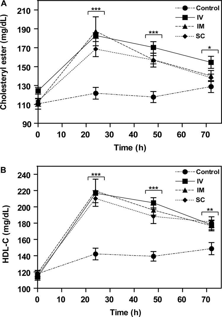
Fig. 4.

Effect of route of administration of rLCAT on mouse lipids. rLCAT (4200 units) was injected intravenously into the tail vein (squares), intramuscularly into hind limb (triangles) or subcutaneously (diamonds) into the back of hapoA-I-Tg mice, and then plasma was removed at the indicated times and analyzed for total cholesterol (A) and HDL-C (B). The control group was treated subcutaneously with saline (circles). Data represent mean ± S.D. (n = 10). *, p < 0.005; **, p < 0.001; and ***, p < 0.0001, experimental versus control group.
