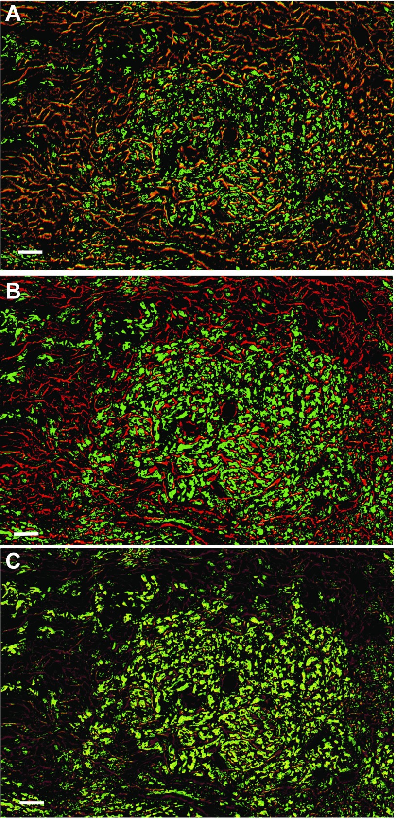
Fig. 7.
Cav1.2 and Cav3.1 are coexpressed in the SA node. FISH on Cav1.2, Cav3.1, and anti-GAP-43 antibody was performed as described in materials and methods. Bars, 25 μm. A: GAP-43 (red) overlaid with Cav1.2 (green) shows considerable expression of Cav1.2 both in the SA node and in the surrounding neurons. B: GAP-43 with Cav3.1 shows that the Ca2+ channel is excluded from the neurons but is expressed in the central nodal area. C: Cav1.2 with Cav3.1 shows that the channels are expressed in the same cells in the central SA nodal tissue.

