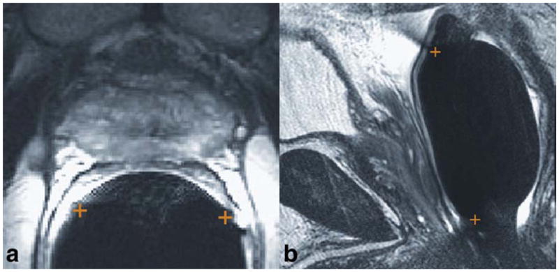
Figure 1.

Identification of the coil location and rotation on (a) axial and (b) sagittal T2-weighted images. The two ends of the coil are marked with +’s. On the axial image, the locations are identified by the indentations in the rectal wall. On the sagittal image, the locations are identified by the dark streaks that emanate anteriorly and outward from the rectal wall at the superior and inferior ends of the balloon-inflated probe. [Color figure can be viewed in the online issue, which is available at wileyonlinelibrary.com.]
