
Figure 3.
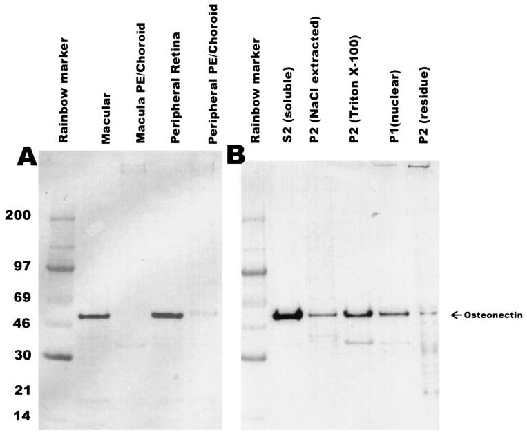
Western blot analyses of osteonectin/SPARC in monkey retinal tissues. Each lane was loaded with approximately 20 μg total protein. (A) Results of Western blot analysis of the macular and peripheral retina punches (same as Fig. 1) probed with rabbit anti-osteonectin/SPARC antibody (1:3000). (B) Results of Western blot analysis of the subcellularly fractionated whole monkey retina. The blots were processed as described in the Methods section. PE, pigment epithelium.