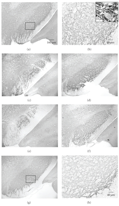
Figure 6.
VGAT immunolabeling in SN in coronal midbrain section of rats after 12.5 minutes of global ischemia at day 56. (a) VGAT-ir in sham rat. (b) Higher magnification of boxed area in (a). The insertion with hematoxylin counterstaining presents a higher magnification of VGAT-ir. Immunohistochemical analysis shows abundant puncta of VGAT-positive dots, and some of these puncta encircle an unlabeled neuron's body and its dendrites. (c)–(g): VGAT-ir in 5 ischemic rats at day 56 after injury. (h): higher magnification of boxed area in (g) shows lower density and less amount of VGAT puncta and fiber network than sham rat.

