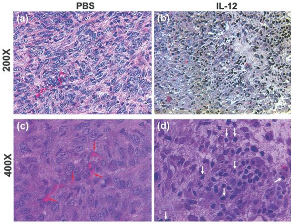
Figure 2.
Angiogenesis and lymphocyte infiltration in primary 4T1 tumors. Parafin-embedded tumor sections were stained with H&E and microscopically examined for blood vessels and tumor infiltrating lymphocytes at 200× (a,b), and 400× (c,d) magnification. Shown for each group is 1 representative slide of 3 tumor samples randomly picked from the 2 experimental groups. Red arrows, blood vessels; white arrows, TILs.

