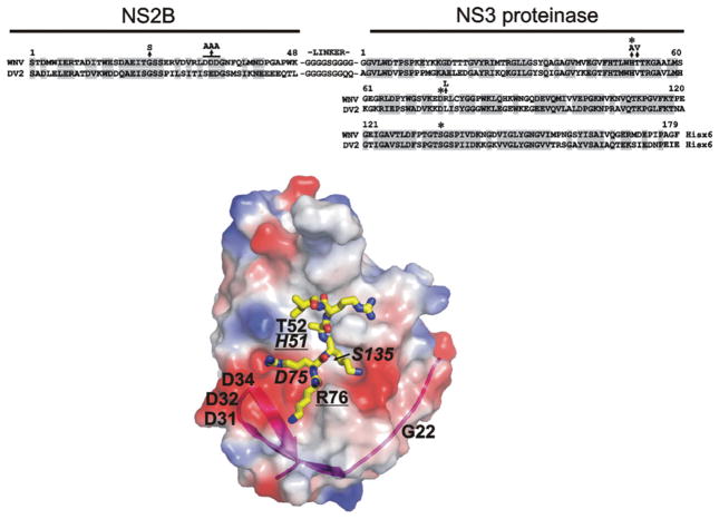
Figure 1. Structure and sequence alignment of the NS2B cofactor and the NS3 proteinase domain of WNV and DV2.
Upper panel: sequence alignment of the NS2B–NS3pro constructs; homologous amino-acid-residue positions are shaded. The asterisks above the sequences indicate His51, Asp75 and Ser135 of the catalytic triad. The arrows indicate mutations (G22S, DDD/AAA, H51A, T52V and R76L). The G22S and DDD/AAA constructs also contained the K48A mutation that inactivated the Lys48↓Gly autolytic cleavage site where glycine is the N-terminal residue of the linker. Lower panel: the structure of WNV NS2B–NS3pro (PDB accession code 2IJO) [8]. The original position of the His51, Asp75 and Ser135 of the catalytic triad (italicized) and the mutant positions are shown. The mutations that affect the binding of the proteinase with the antibodies are underlined. The surface is coloured by electrostatic potential (negative, red; positive, blue). The magenta ribbon shows NS2B contributing to the NS3pro active site. The positioning of a substrate-mimetic peptide (KRKARI) is shown in yellow. The model was presented using PyMol software (DeLano Scientific; http://www.pymol.org).

