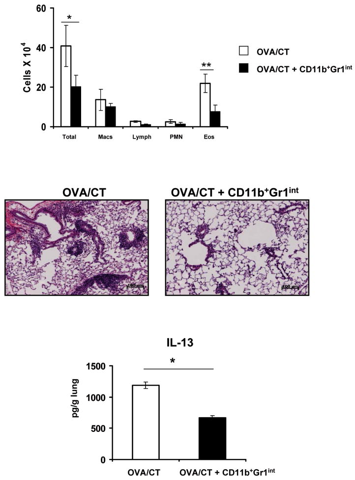
Figure 7.
CD11b+Gr1int cell-mediated prevention and treatment of eosinophilic airway inflammation in vivo. Mice were antigen-sensitized by three daily consecutive intranasal treatments with OVA plus cholera toxin (CT) followed by 5 d of rest. CD11b+Gr1int cells were generated from bone-marrow progenitor cells in the presence of GM-CSF (10ng/ml) and LPS (1μg/ml) and then adoptively transferred intratracheally (1 × 10⁶ cells/mouse) into mice that had received OVA/CT. Control mice did not receive any cells. Mice were then challenged with aerosolized OVA daily for 7 d. Total and differential cell counts in the BAL fluid (upper panel) were enumerated. Values are mean ± SEM *, P<0.05 and **, P<0.01. H&E staining (middle panel) of lung sections was performed. Lung infiltrates around bronchovascular bundles were of +5 grade in animals that did not receive CD11b+Gr1int cells and +1 grade in those that did. IL-13 present in lung homogenates (lower panel) was measured by ELISA and presented as the mean value ± SEM *, P < 0.05.

