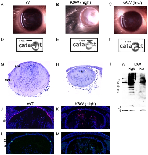
Figure 2. K6 on Ub is essential for proper lens formation and clarity.
Slit-lamp photographs of P90 mouse lenses. Lenses expressing high levels of K6W-Ub (A) show severe cataracts whereas lenses from animals expressing low levels of K6W-Ub (C) are clear, comparable to wild type (B). (D, E, F) Head-on photographs of P30 lenses. Lenses expressing high levels of K6W-Ub are cloudy and opaque. Note that the print behind the lens in panel E cannot be seen as it is in panels D and F. Lens from animals expressing low levels of K6W-Ub are clear comparable to Wt. (G, H) Light micrographs from E18.5 days Wt and K6W-Ub lenses. Lenses expressing high levels of K6W-Ub were ∼2/3 the size of Wt lenses. (I) P30 lenses from high or low K6W-Ub-expressing animals show different levels of K6W-Ub-containing conjugates. Lenses from Wt and transgenic animals were lysed and expression of transgene was determined by western blotting using anti-RGS(His)4. (J–M) Fluorescent micrographs of E18.5 K6W-Ub-expressing lens show attenuated proliferation compared to Wt. (J, K) BrdU (red) incorporation assay was used to detect S-phase cells in mouse lenses. K6W-Ub-expressing lenses show limited incorporation of BrdU compared to Wt. (L, M) Phospho-H3 (green), also shows that K6W-Ub-expressing lenses have decreased proliferation compared to Wt lenses. Immunohistochemistry was used to detect incorporation of BrdU and expression of phospho-H3, using anti-BrdU and anti-phospho-H3 antibodies respectively. DAPI was used to stain nuclei.

