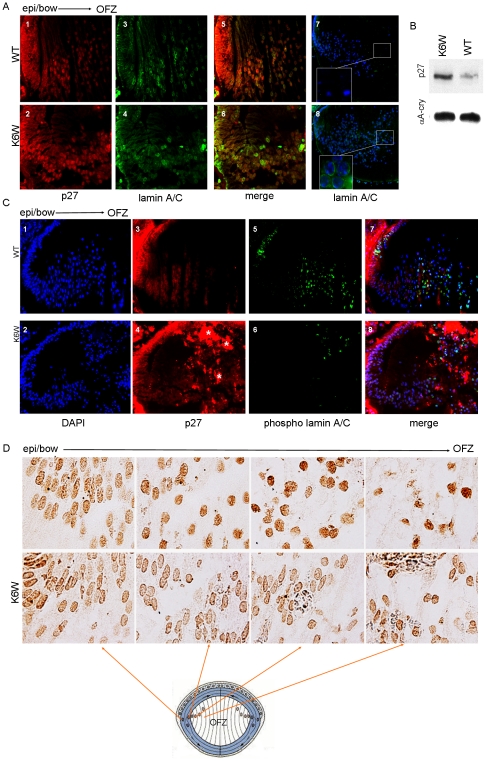
Figure 4. K6 on Ub is required to direct lens denucleation.
(A) Fluorescent micrographs of E18.5 K6W-Ub-expressing (panel 2, 4, 6, 8) and Wt (panel 1,3, 5, 7) lenses show distribution of p27 (red) and lamin A/C (green). In Wt lenses p27 is localized at the bow region of the lens and is concentrated in nuclei, but is also present in the cytoplasmic compartments of lens fiber cells. There is a gradual decrease in p27 expression from the bow region to the OFZ. Transgenic lenses (panel 2) show greater retention of p27 (note elevated red staining) in nuclei and the fiber mass as compared to Wt (panel 1). Intact nuclear membranes in the core of the lens are only seen in transgenic lenses (panel 8). While in Wt, no nuclear membranes are apparent and only DNA fragments are observed in the core of the lens (panel 7). (B) P1 K6W-Ub transgenic lenses show stabilization of p27 protein. Lenses were lysed and levels of endogenous p27 were determined by western blotting using anti-p27 antibody. Equal loads are shown by western blotting using anti-αA crystallin antibody. (C) Fluorescent micrographs of E18.5 K6W-Ub-expressing (panel 4) and Wt (panel 3) lenses show distribution of p27 (red) and phosphorylated lamin A/C (green) (panels 6 and 5). Localization of p27 is the same as in (A, panel 1 and 2). Asterisks show non-specific staining (panel 4). In addition, lenses that express K6W-Ub (panel 6) show decreased levels of phosphorylated lamin A/C (green) toward the lens core as compared to Wt (panel 5). Immunohistochemistry was done to detect levels of p27, phosphorylated lamin A/C, lamin A/C using anti-p27, anti-lamin A/C (phospho Ser 392) and anti-lamin A/C, respectively. DAPI was used to stain nuclei. Note that phosphor-lamin is detected more in the dividing epithelia of Wt lenses rather than transgenic lenses. (D) Light micrographs of P2 mice Wt and K6W-Ub transgenic lenses show the distribution of DNAse IIβ (dark brown). Comparable sections from the bow region to the edge of the OFZ of the lens. K6W-Ub-expressing lenses show accumulation of DNAse IIβ around the nuclear envelope for all regions. Going from the bow towards the OFZ, more DNAse IIβ enters the nucleus and less accumulates at the nuclear envelope. Immunohistochemistry was done to detect distribution of DNAse IIβ using anti-DNAse IIβ antibody.

