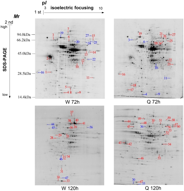
Figure 1. Proteome map of honeybee worker and queen larvae at developmental stages 72 hours (w 72 h and Q 72 h) and 120 hours (W 120 h and Q 120 h).
2-DE separation was performed on IPG gel strips (17 cm, 3- 10L) followed by SDS-PAGE on a vertical slab gel (12%). Protein spots of known identity are marked with color codes, red indicating upregulated proteins and blue indicating down regulated.

