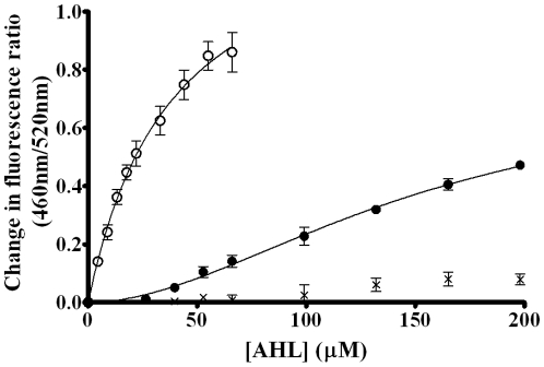
Figure 7. Binding profiles of the interactions of AHLs with Lymphocyte membranes.
Binding profiles of 3-oxo-C14-HSL (Δ, hyperbolic), 3-oxo-C12-HSL (•, sigmoidal) and 3-oxo-C10-HSL (×, neither) on titration to di-8-ANEPPS labeled T-Lymphocytes (40,000 cells/ml) at 37°C normalised to DMSO controls. Profiles were fitted to simple hyperbolic and sigmoidal binding models (equations 1 and 2) and F-Tests were used to determine the best fitting model. In each experiment n = 3, ±SEM.

