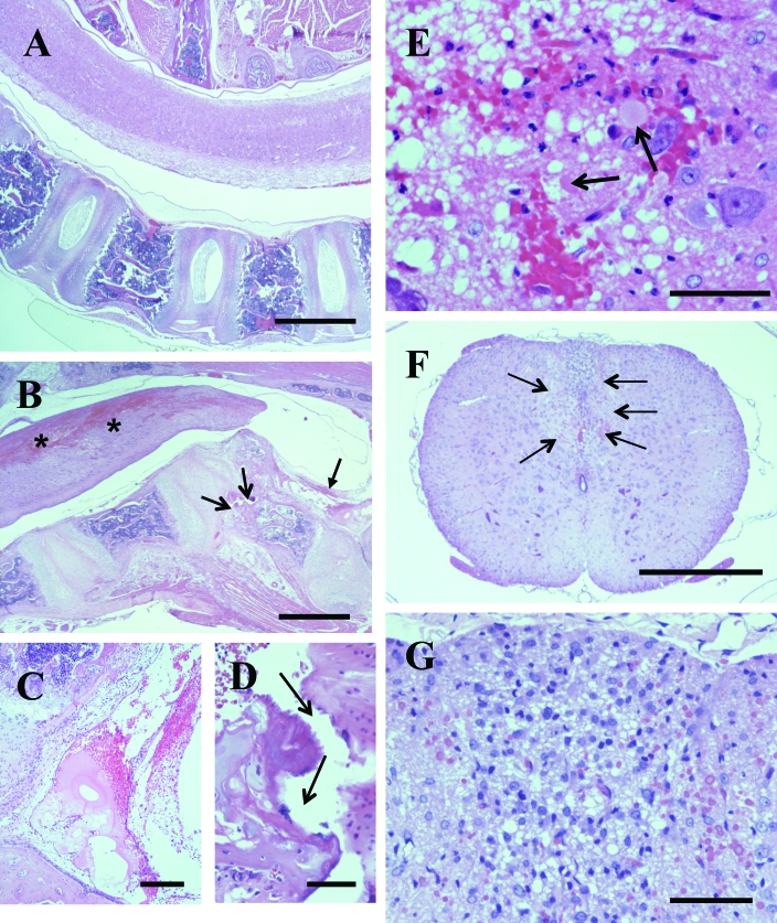
Figure 1.
Spine with spinal cord in myelin-deficient (md) rat. (A) Sagittal section of the spine including intact spinal cord. Bar, 1000 μm. (B) Vertebral canal is markedly narrowed over the angular protrusion of a vertebrae with its body fractured (open arrows). There is marked fibrinohemorrhagic exudate in the adjacent area of the vertebral canal (solid arrow). The spinal cord has large interconnected hemorrhages predominantly in the dorsal area (asterices). Bar, 1000 μm. (C) Higher magnification of the fibrinohemorrhagic exudate in the lumen of the vertebral canal indicated by the solid arrow in panel B. Bar, 200 μm. (D) Higher magnification of the fracture in the vertebral body (open arrows) in panel B. Bar, 50 μm. (E) Histologic detail of the area with hemorrhage in the spinal cord in panel B. There is patchy hemorrhage, marked vacuolation of the neuropil, scattered swollen axons (arrows), and scattered neutrophils. Bar, 50 μm. (F) Cross section of the thoracic spinal cord, with a large hypercellular area in the dorsal column (arrows). Bar, 500 μm. (G) Higher magnification of the area of hypercellularity in the dorsal column in panel F, showing diffuse vacuolation of neuropil and infiltration by mononuclear cells, presumably macrophages. Bar, 50 μm.

