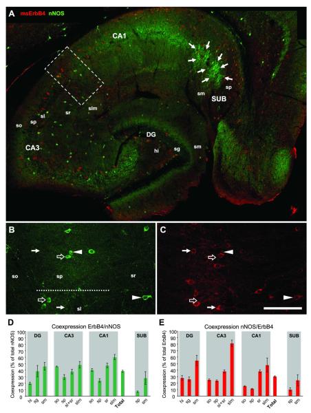
Figure 5. One third of nNOS-positive interneurons express ErbB4.
nNOS-immunoreactive somata are located in all hippocampal areas, with the highest densities in hi, and in sp of CA1-3 and SUB (A). Immunoreactivity for nNOS is evident on neurites in the inner third of sm in DG, and in all layers of CA1. The highest density of nNOS-ErbB4 coexpressing cells is in slm of CA3. Note the cluster of strongly immunoreactive pyramidal cells in SUB (arrows in A) that are consistently negative for ErbB4. Single channel imaging in CA1-3 (B-C) shows cells that express either nNOS (open arrows) or ErbB4 (arrows), as well as double-immunoreactive somata (arrowheads). Regional analysis of coexpression (n=3) in all nNOS-positive cells (D) and all ErbB4-positive cells (E). Please note that “Total” represents only coexpression in DG and CA1-3; in order to focus on interneurons SUB was excluded to avoid bias towards the high number of nNOS-positive/ErbB4-negative pyramidal cells in sp of SUB. Abbreviations: see Fig.2. Scale bar = 290μm (A), 100μm (B,C).

