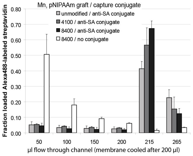
Figure 4.
Anti-streptavidin antibody conjugate-bound Alexa 488-labeled streptavidin (SA) captured at membranes during heated flow (50 μl per minute) and released with cooled flow (50 μl per minute). Each 50 μl sample contained 10 nM streptavidin with either no antibody conjugate (white), or 100 nM anti-streptavidin pNIPAAm conjugate (grey to black). Membranes tested were unmodified Loprodyne (light grey), 4,100 Mn-graft (medium grey), or 8,400 Mn-graft (black and white) pNIPAAm-polymerized Loprodyne. Fluorescence of recovered flow fractions was measured using a plate reader fluorimeter against a standard curve.

