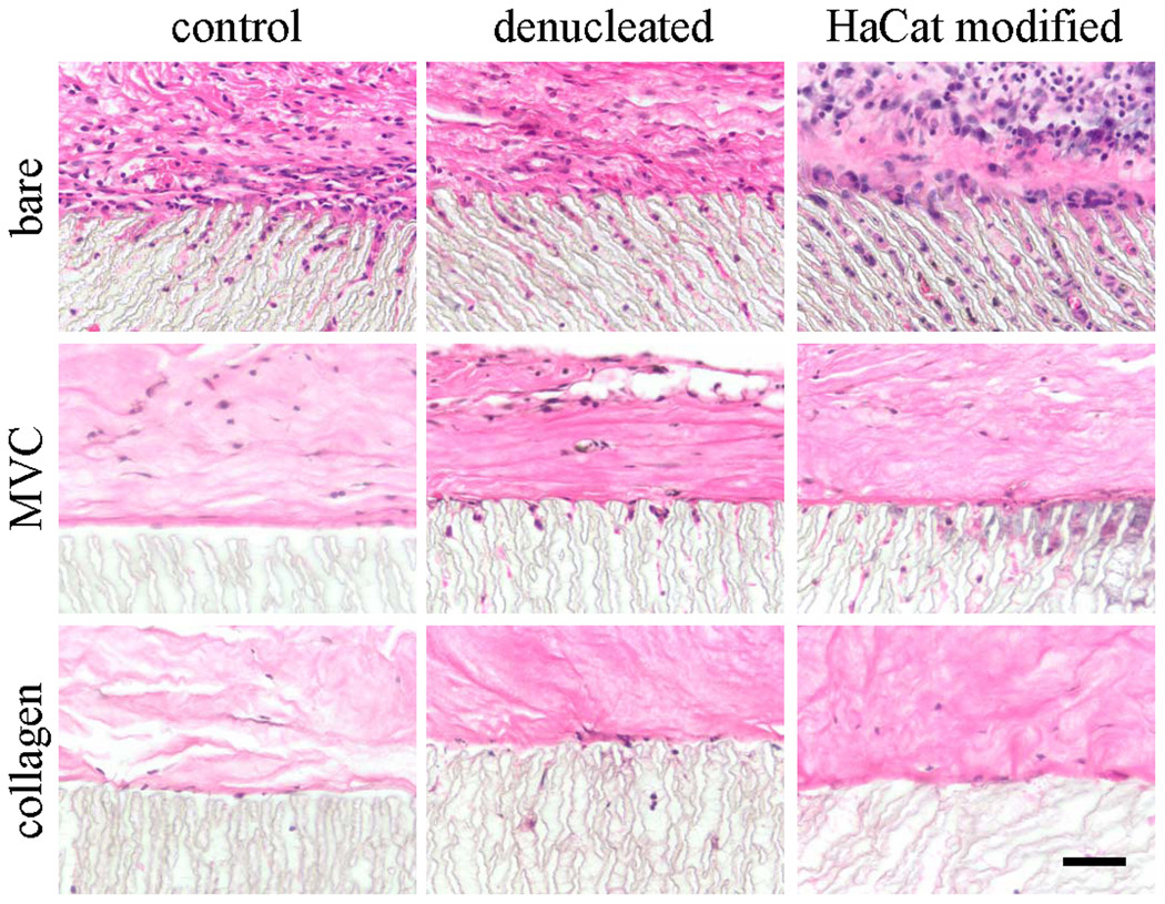
Figure 1.

Light micrographs of hematoxylin and eosin-stained paraffin sections of ePTFE discs implanted for 28 days as is (bare) or embedded in a microvascular construct (MVC) or an avascular collagen gel (collagen). EPTFE discs were used without modification (control), denucleated, or modified with a secreted extracellular matrix preparation (HaCaT modified). Scale bar = 50 microns.
