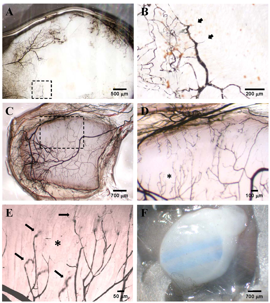
Figure 5.

Vascular India ink casts of ePTFE discs implanted for 28 days used to visualize the perfusion-competent microvessel network in control bare ePTFE (A, B), control ePTFE embedded in a microvascular construct (C–E), or control ePTFE embedded in collagen alone (F). Panel B, a higher magnification of the area marked by the dotted box in panel A, shows short vessel branches that superficially enter the ePTFE interstices (arrow heads). The dotted box in panel C marks the area magnified and shown in panel D. The asterisk in panel D marks the area further magnified to better visualize perfused vessel ends plunging into the ePTFE interstices (arrows). The parallel blue stripes in panel F are markings printed onto the ePTFE by the manufacturer which are visible through the collagen gel.
