
Figure 4.
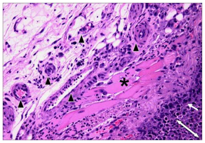
Hematoxylin and eosin stained section (at x 200 magnification) of KHT sarcoma treated with OXi4503 (25 mg/kg). The post-treatment necrotic tumor is defined by the double-headed arrow while the two- or three-cell layer of the viable tumor rim is shown with a single-headed arrow. Preserved muscle (*) and blood vessels (black arrowheads) are clearly evident in the surrounding, unaffected tissue (Siemann DW, unpublished results).