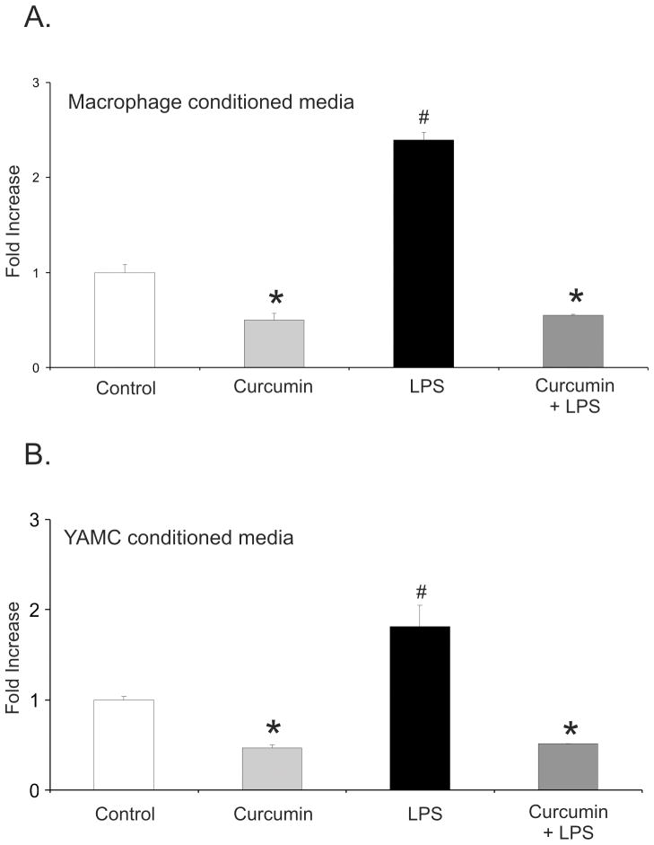
Figure 3.
Chemotaxis Assay with calcein red-orange AM stained bone marrow-derived mouse neutrophils against conditioned medium obtained from intraperitoneal macrophages (A) or YAMC cells (B) treated with DMSO (control), 50 μM curcumin, LPS (10 ng/mL), or LPS with curcumin (the same treatment conditions as described in Fig. 1). *Statistically significant differences (p≤0.05) between values from cells treated with curcumin or curcumin/LPS and control or LPS treatment; # statistically significant differences between values from cells treated with LPS alone and other respective treatments (ANOVA followed by Fisher PLSD post-hoc test).

