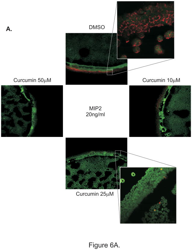
Figure 6.
Under agarose neutrophil migration assay: F-actin was labeled after migration with Alexa-647 conjugated phalloidin and nuclei were counterstained with sytox green and cells were imaged under a confocal laser scanning microscope. (A) Control DMSO-treated PMN or PMN pretreated with 10–50 μM of curcumin for 30 min prior to the assay were loaded into different wells cut in 1.6% agarose gel. Recombinant MIP-2 (20ng/ml) was placed in the central well. Each well was placed at equal distance from each other. High magnification images from PMN treated with DMSO or 25 μM curcumin are depicted as crop-outs. (B) Curcumin was homogeneously added to the gel at 25 or 50 μM. PMN or recombinant MIP-2 (20ng/ml) were loaded into adjacent, evenly spaced wells.


