Figure 7.
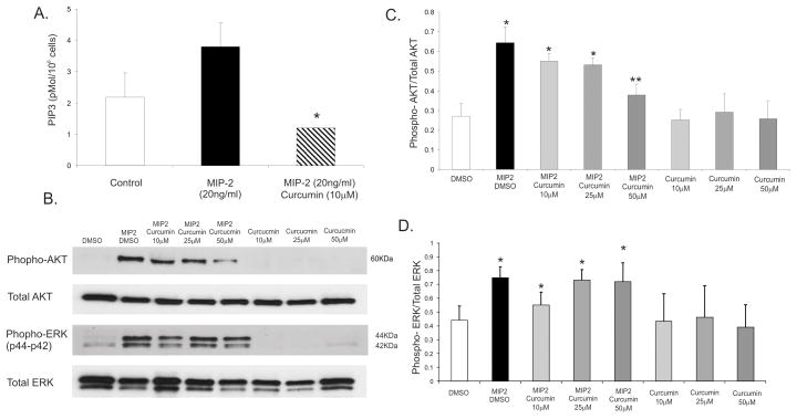
(A) ELISA for PIP3 concentration (pMol/106 cells) in bone marrow derived-neutrophil treated with 10–50μM of curcumin 5 minutes prior to a 30-second stimulation with MIP-2 (20ng/ml). (B) Western blot analysis of phospho-AKT (Ser473) or to phospho-p44/42 MAPK (Thr202/Tyr204) in PMNs treated with 10–50μM of curcumin 5 minutes prior to a 30-second stimulation with MIP-2 (20ng/ml). Total AKT and total ERK were evaluated as loading control. * p≤0.05 curcumin/MIP-2 vs. control or MIP-2 alone (ANOVA followed by Fisher PLSD post-hoc test). (C) Densitometric analysis of AKT and (D) ERK phosphorylation in response to MIP-2 with or without 10–50μM curcumin.

