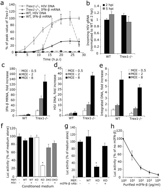
Fig. 3. Cytosolic HIV DNA in Trex1−/− cells is the trigger for IFN expression.
(a) Cytosolic HIV DNA accumulates at higher levels early after infection in Trex1−/− (KO) than in WT primary MEFs. IFN-β expression lags behind DNA build up and only occurs in Trex1−/− cells. HIV DNA and IFN-β mRNA were measured by qPCR and qRT-PCR, respectively. (b) Trex1 deficiency does not affect viral entry. Comparable numbers of virions enter WT and Trex1−/− cells as determined by qRT-PCR of HIV genomic RNA (gRNA) assessed 2 and 5 hpi. (c-e) Increasing multiplicity of infection (MOI) leads to more cytosolic HIV DNA accumulation and IFN-β expression in Trex1−/− MEF, but not to more chromosomal integration, compared to WT MEF. WT and Trex1−/− MEFs were infected with HIV at indicated MOI. IFN-β mRNA (c) and cytosolic HIV DNA (d) were measured as in (a). Integrated DNA (e) was measured by a two-step semi-quantitive PCR assay (8, see ONLINE METHODS). (f) Conditioned medium from Trex1−/− MEF, but not from WT and Trex1−/−Irf3−/− (DKO) MEF, inhibits de novo HIV infection. Conditioned medium, obtained from cells that were either uninfected or infected with HIV-GFP, was added to WT MEFs that were then infected with HIV-Luc. Luc activity was measured 48 hpi. (g) IFN-β contributes to most of the antiviral activity secreted by Trex1−/− MEF. Conditioned medium was pre-incubated with or without mouse IFN-β neutralizing antibody before being added to WT MEFs with HIV-Luc. (h) The antiviral effect of varying concentrations of purified recombinant mIFN-β on HIV-Luc infection in WT MEF. *, P <0.01, Student's t-test. Error bars indicate S.D. of three independent experiments.

