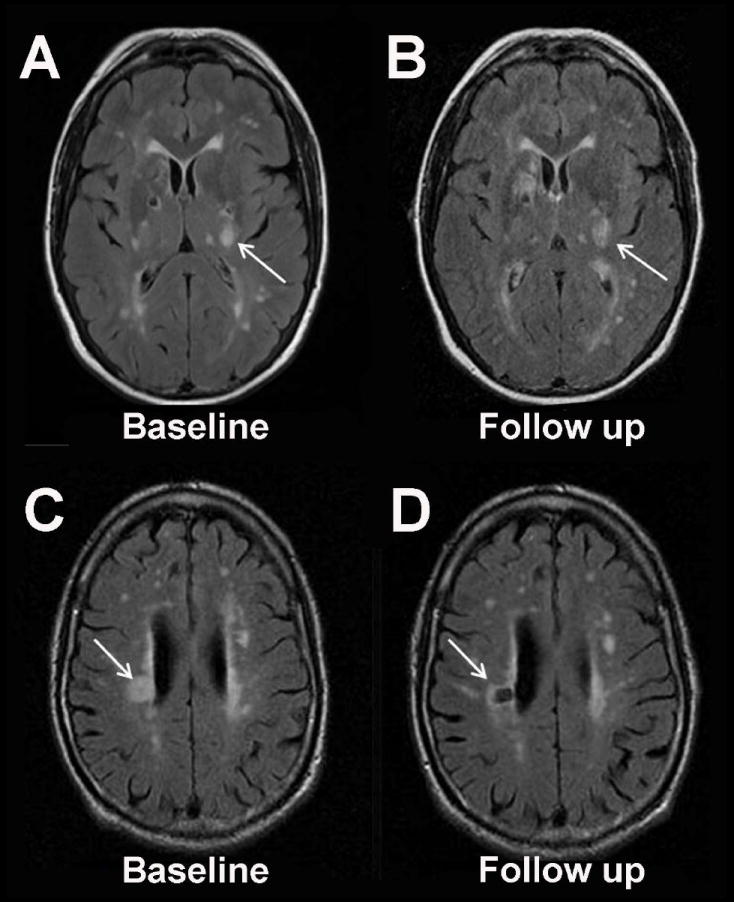
Figure 1. Acute Small Vessel Infarction May Evolve Into White Matter Lesions.

Recent data suggest that the majority of acute small artery infarctions on MRI evolve into T2-hyperintense white matter lesions (Panels A and B, fluid attenuated inversion recovery (FLAIR) MRI at 4 days and 65 days after acute infarction in the left internal capsule) rather than chronic cavitated lesions (Panels C and D, FLAIR MRI at 17 days and 7 months after acute infarction in the right corona radiata).9 This figure was graciously provided by Dr. Gillian M. Potter and Dr. Joanna M. Wardlaw.
