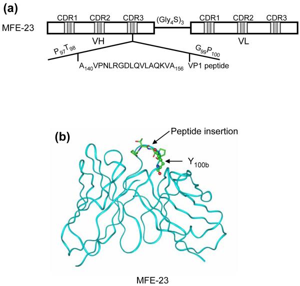
Fig. 1.
Schematic presentation of the construction of B6-1 and B6-2. (a) Insertion of the RGD containing peptide sequence of VP1 (A140 to A156)17 into the CDR3 loop (between T98 and G99) of the VH chain of MFE-23 that gives B6-1. (b) Ribbon diagram of the x-ray structure of MFE-23.25 CDR3 loop residues P97 to P100 of the VH chain of MFE-23 are shown in stick presentation, and the site of peptide insertion in MFE-23 is indicated. Y100b that was mutated to P100b to give B6-2 is shown in ball-and-stick presentation.

