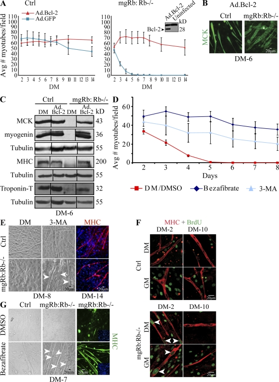
Figure 3.
Rescue of Rb-deficient myotube degeneration by Bcl-2 and autophagy inhibitors. (A) Mean number of myotubes per field in indicated myoblast cultures induced to differentiate after transduction with Ad.GFP or Ad.Bcl-2. Each point represents the mean ± SD of six fields from six independent experiments. Bcl-2–rescued, twitching Rb−/− myotubes are shown in Video 2. (inset) Western blot for Bcl-2 in Ad.Bcl2-transduced versus uninfected primary myoblasts. (B) Immunostaining for MCK (green) in Ad.Bcl-2–transduced control (ctrl) and Rb−/− myotubes at DM-6. (C) Western blot for MCK, myogenin, MHC, and troponin T in indicated cultures at DM-6 after transduction with Ad.Bcl-2. Tubulin was used as a loading control. Black lines indicate that intervening lanes have been spliced out. (D) Mean number of Rb−/− myotubes after 3-MA treatment, bezafibrate, or DMSO/vehicle as indicated. Counts are mean ± SD of six fields (n = 4). (E, left and middle) Brightfield images of the indicated myotubes treated or not with 3-MA at DM-8. (right) Immunostaining for MHC (red) at DM-14 in the presence of 3-MA. Arrowheads label 3-MA–rescued Rb−/− myotubes at DM-8. 3-MA–rescued, twitching Rb−/− myotubes are shown in Video 4. (F) Immunostaining for BrdU (green) and MHC (red) of 3-MA–treated cultures at DM-2 or DM-10. Rb−/− myotubes incorporated BrdU at DM-2 (arrowheads) but not DM-10. (G) Brightfield images (left and middle) and MHC staining (right) of the indicated cultures treated or not with bezafibrate. Arrowheads point to bezafibrate-rescued Rb−/− myotubes.

