Figure 3.
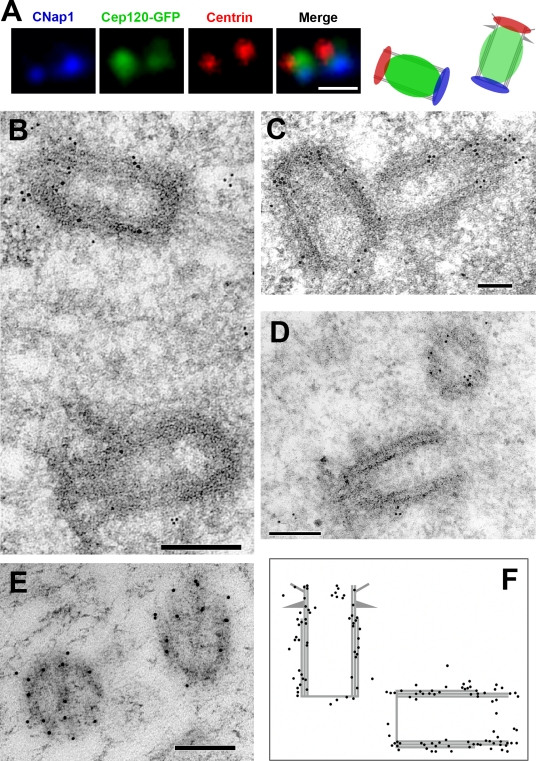
Cep120 is associated with the outer wall of centrioles. (A) Immunofluorescence localization of Cep120 relative to centriole markers. G1-phase RPE-1 cells expressing Cep120-GFP were fixed and stained for centrin to mark the distal end of centrioles (red), C-Nap-1 to mark the proximal end of centrioles (blue), and Cep120-GFP (green). The panel on the right is a representation of the observed spatial distribution. (B–E) Immunoelectron microscopy localization of Cep120 on centrioles. NIH3T3 cells were fixed and processed for transmission electron microscopy and incubated with anti-Cep120 antibodies followed by 10-nm gold-conjugated secondary antibodies. (F) Schematic of observed gold particle distribution (130 particles). Bars: (A) 0.5 µm; (B, C, and E) 100 nm; (D) 200 nm.

