Figure 7.
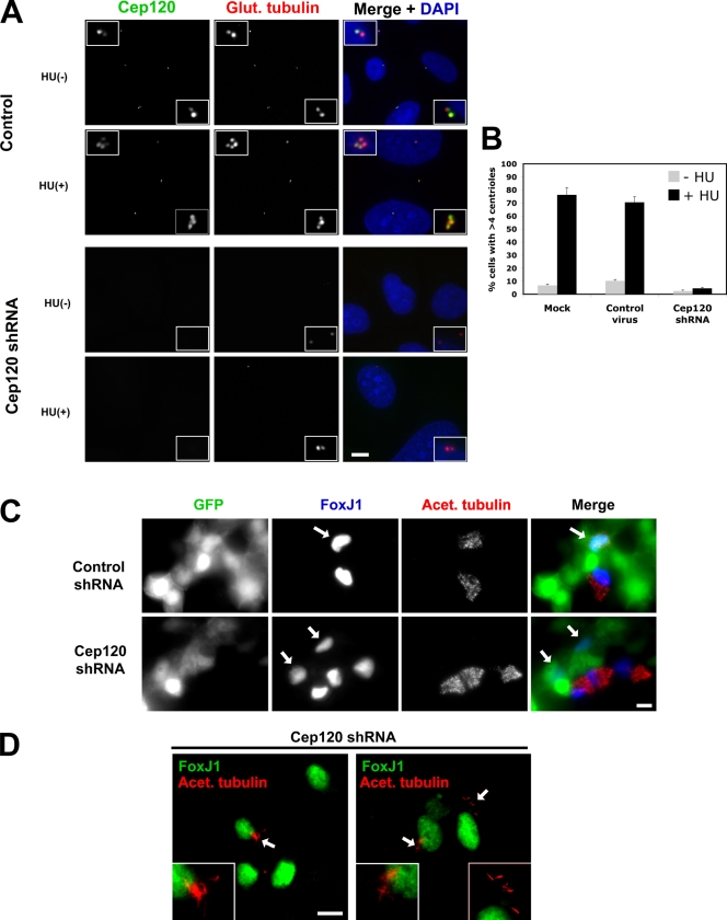
Cep120 is required for centriole amplification. (A) Block of centriole overduplication in Cep120-depleted cells. U2OS cells were infected with lentivirus expressing shRNA targeting Cep120 or a control shRNA, arrested in S phase by hydroxyurea (HU) treatment for 48 h, and stained for Cep120 (green), glutamylated tubulin (red), and DNA (blue). (B) Quantification of centriole overduplication in Cep120 shRNA, control shRNA, or mock-infected U2OS cells. Results shown are a mean of two independent experiments (n = 400 for each sample). (C) Block of centriole and cilia assembly in Cep120-depleted MTECs. MTECs were infected with Cep120 shRNA or control shRNA lentivirus 2 d before establishing ALI and then were stained at ALI+8 for GFP to mark infected cells (green), FoxJ1 to identify cells committed to the ciliated cell fate (blue), and acetylated tubulin (red). Arrows indicate cells infected with shRNA-expressing lentivirus. (D) Examples of cilia formation in MTECs with partial centriole duplication defects. Cells were stained for FoxJ1 (green) and acetylated tubulin (red; arrows mark regions magnified in insets). Insets are magnified images of the centrosome region. Data are means ± SD. Bars, 10 µm.

