Fig. 2.
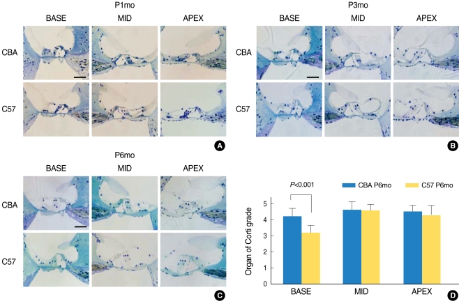
Serial light microscopy changes of the organ of Corti from CBA (upper panels) versus C57 (lower panels) mice from base, mid, and apical regions of the cochlea. At P1mo (A), the morphology of the organ of Corti did not show differences between CBA and C57 strains. However, from P3mo (B), C57 mice showed degeneration of outer hair cells at the base of the cochlea. At P6mo (C), C57 mice consistently showed more severe damage in the region of the outer hair cells, compared with CBA. A rank-order grading method was used to rate the condition of supporting cells and the general shape of the organ of Corti. The average value for regional supporting cell condition in the base of C57 mice was significantly lower than that of CBA mice at P6mo. (D) Six mid-modiolar sections were counted and averaged to obtain a value from one animal. Scale bar 50 µm.

