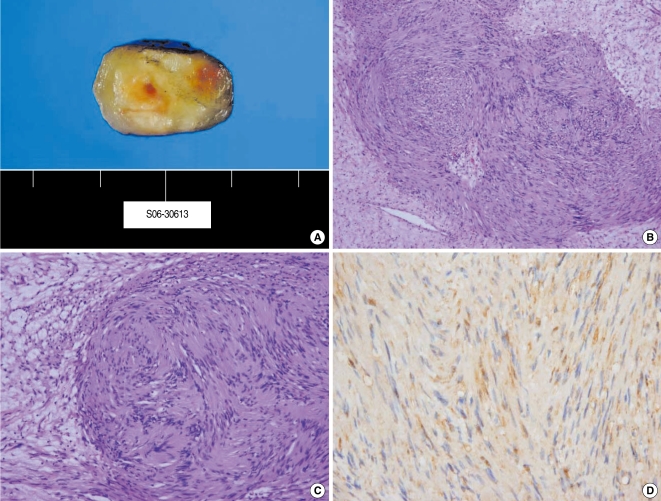
Fig. 4.
The histologic findings of the schwannoma in case 2. The mass had a yellowish color and it had a well capsulated sursolid form (A). The schwannoma cells were organized in compact bundles with the nuclei arranged in a palisade manner (Antoni type A) and the cells were partly dispersed with loose reticular fibers (Antoni type B) (B: H&E, ×100; C: H&E, ×400). The schwannoma cells showed immunoreactivity for S-100 protein (D: Immunohistochemistry).

