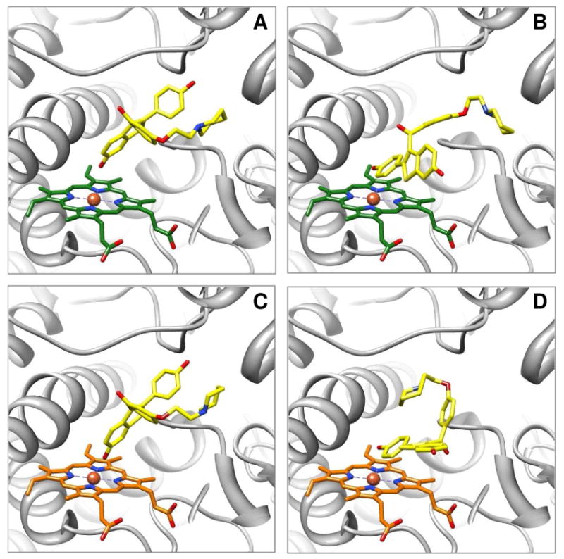
Figure 2.

Molecular models of raloxifene in the active site of CYP3A4 predicted by AutoDock 3.0. Raloxifene was docked into 1W0E, with an unmodified heme, or into 1W0E_Modified, where partial charges were assigned to the heme. A representative conformation from the largest/lowest energy cluster, predicting dehydrogenation, from docking with 1W0E is depicted in (A), and from docking with 1W0E_Modified is depicted in (C). The dehydrogenation orientation of raloxifene is portrayed in (A) and (C), with the hydroxyl group on C-6 of the benzothiophene moiety oriented toward the heme and close to the Fe. A representative conformation predicting raloxifene 3′ hydroxylation from docking with 1W0E is depicted in (B), and from docking with 1W0E_Modified is depicted in (D). 3′ hydroxylation of raloxifene is portrayed in (B) and (D), with the hydroxyl group on C-4′ of the phenol moiety oriented toward the heme. CYP3A4 is shown in a ribbon format, iron as a sphere, heme (green for 1W0E and orange for 1W0E_Modified) and raloxifene (yellow) in color-coded sticks: nitrogen = blue, oxygen = red. Molecular graphics images were produced using the UCSF Chimera package from the Resource for Biocomputing, Visualization, and Informatics at the University of California, San Francisco.
