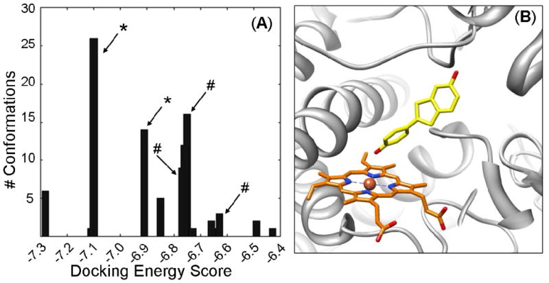
Figure 3.

Docking of a simplified benzothiophene molecule into the CYP3A4 active site. (A) Histogram of 100 conformations from docking 2-(4-hydroxy-phenyl)-benzothiophene-6-ol into 1W0E_Modified (with heme partial charges assigned) using AutoDock 3.0. Conformations generated by AutoDock were clustered with an RMSD of 2 Å, and plotted by the lowest energy conformation of each cluster. (*) Indicates clusters with the phenol moiety oriented toward the heme. (#) Indicates cluster with the benzothiophene moiety oriented toward the heme. Unmarked clusters have “inactive” conformations. (B) A representative conformation from the most abundant/lowest energy cluster of 2-(4-hydroxy-phenyl)-benzothiophene-6-ol, which predicts that the phenol moiety is oriented toward the heme. CYP3A4 is shown in a ribbon format, iron as a sphere, heme modified with partial charges (orange) and docking 2-(4-hydroxy-phenyl)-benzothiophene-6-ol (yellow) in color-coded sticks: nitrogen = blue, oxygen = red. Molecular graphics images were produced using the UCSF Chimera package from the Resource for Biocomputing, Visualization, and Informatics at the University of California, San Francisco.
