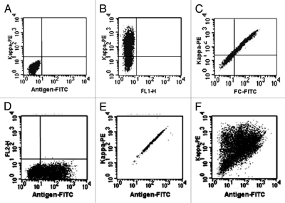
Figure 4.
Flow cytometry analysis of antibodies displayed on the cell surface. Flp-In 293 cells were transiently transfected by FVTM containing the same human antibody gene (B–E) or a library of antibody genes (F). Cells were labeled 48-hrs post transfection with fluorescence-conjugated antibodies and/or antigen and then analyzed by flow cytometry. Kappa-PE, Phycoerythrin (PE)-conjugated mouse anti-human Kappa chain antibody; FC-FITC, FITC-conjugated mouse anti-human IgG antibody; Antigen-FITC, FITC-conjugated specific antigen. The x- and y-axis indicate the fluorescence intensities of FITC and PE fluorophores respectively. The cells carrying vector without antibody genes were used as the control (A).

