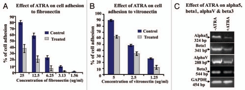
Figure 4.
(A) MDA-MB-231 cells grown in presence (Treated) and in absence (Control) of 20 µM ATRA were allowed to bind with different concentration of fibronectin (25 µg/ml, 12.5 µg/ml, 6.25 µg/ml, 3.125 µg/ml, 1.56 µg/ml) coated in 96 well plate. After 1.5 h incubation at 37°C wells were washed and cells were trypsinized. Numbers of bound cells were counted on a haemocytometer slide and % of adhesion was calculated. (B) MDA-MB-231 cells grown in presence (Treated) and in absence (Control) of 20 µM ATRA were allowed to bind with different concentration of vitronectin (5 µg/ml, 2.5 µg/ml, 1.25 µg/ml) coated in 96-well plate. After 1.5 h incubation at 37°C wells were washed and cells were trypsinized. Numbers of bound cells were counted on a haemocytometer slide and % of adhesion was calculated. (C) RT-PCR was performed in control (lane −ATRA) and ATRA treated (lane +ATRA) MDA-MB-231 cells with α5, β1, αv and β3 primer. GAPDH was used to confirm total RNA integrity and equal loading.

