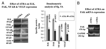
Figure 5.
(A) Western blots were performed in MDA-MB-231 cells grown in absence (lane −ATRA) and in presence (lane +ATRA) of 20 µM ATRA for 48 h as described in methods. Membranes were developed using anti-ILK, anti-FAK, anti-NFκB and anti-VEGF primary antibody, keeping actin as internal control. (B) RT-PCR of FAK was performed in MDA-MB-231 cells grown without (lane −ATRA) or with (lane +ATRA) 20 µM ATRA. PCR products were run on 2% agarose gel to visualize the bands.

