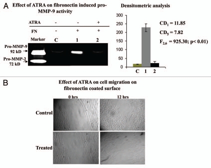
Figure 8.
(A) MDA-MB-231 cells (300,000 cells/1.5 ml) grown in presence (lane 2) and in absence (lane 1) of 20 µM ATRA for 48 hrs. Both control and ATRA treated cells were then allowed to grow in presence of 20 µg/ml fibronectin for 2 h in SFCM. Lane C represents MDA-MB-231 cells grown in absence of fibronectin as well as ATRA. SFCMs were then subjected to gelatine zymography as before. Lane M is the marker lane, showing pro-MMP-9 and pro-MMP-2 activity in the SFCM, collected from HT-1080 cells. (B) Cell migration efficiency of Control and ATRA treated MDA-MB-231 cells were observed under inverted microscope by creating wounds in cell culture dishes. Cells were allowed to grow in presence of fibronectin ECM ligand to observe the efficacy of their migrations in SFCM.

