
Figure 3.
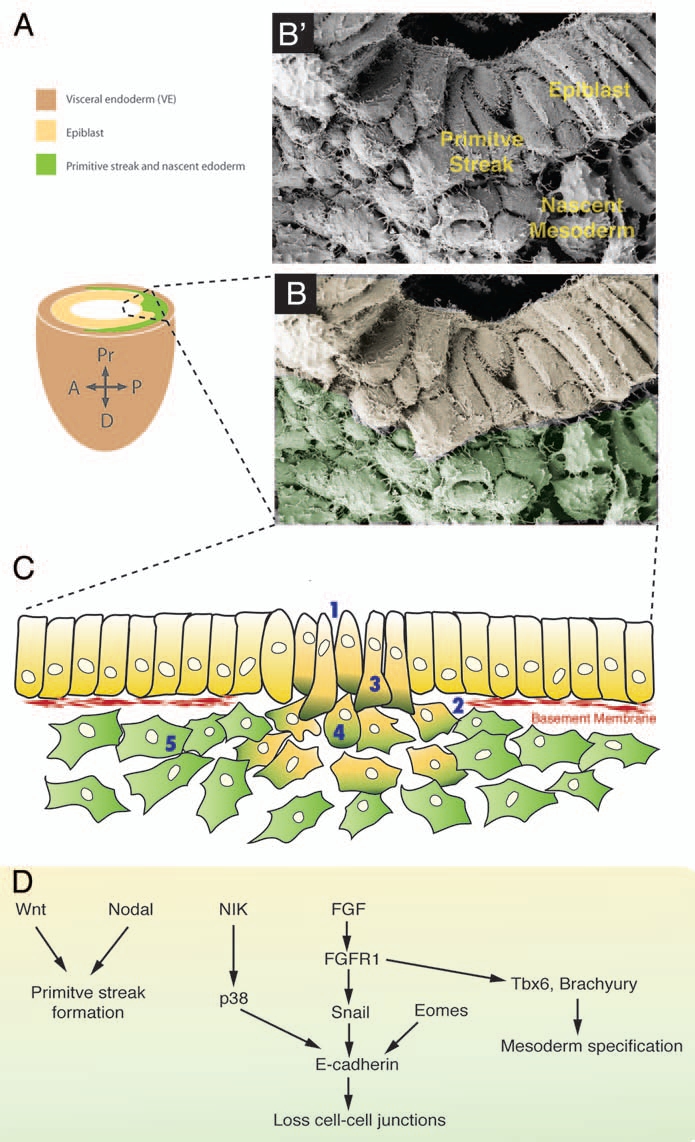
EMT at the mouse primitive streak. (B') Scanning electronic micrograph showing a transverse section through a E7.5 mouse primitive streak. (A) Scheme of the embryonic part of a E7.5 mouse embryo. Dashed box outlines the primitive streak. (B) Scanning micrograph of (A) color-coded for the different germ layers. (C) Cells undergo an eMT event at the primitive streak. (1) First intercellular spaces appear between cells and (2) basal lamina breaks down. (3) Cells acquire a bottle shape, (4) round up as they travel through the streak, and (5) finally acquire a stellate morphology and migrate away from the streak. (D) Signaling pathways that regulate the different EMT steps at the murine primitive streak.