
Figure 2.
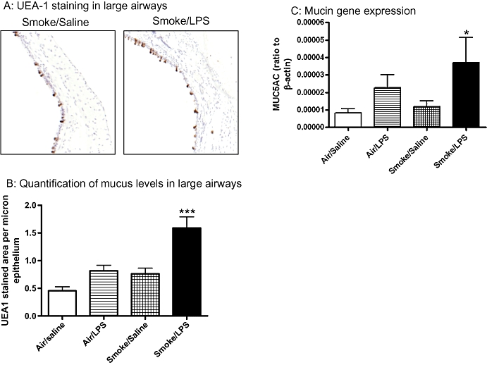
Mucus levels are elevated in response to exposure to a combination of LPS and tobacco smoke. Sprague-Dawley rats were exposed to tobacco smoke or air for 30 min twice a day for 2 days. On the morning of day 3 rats were exposed to saline of LPS (0.3 mg·mL−1) for 30 min followed by tobacco smoke or air 5 h later. Twenty-four hours after LPS exposure the levels of mucus were determined by UEA-1 staining and MUC5AC gene expression. Data shown are (A) representative pictures of UEA-1 staining (B) quantification of UEA-1 stained area expressed as mean and SEM of n= 8 per group and (C) MUC5AC gene expression levels in the lung tissue expressed as mean and SEM of n= 7–8 per group. (*P < 0.05, ***P < 0.001 compared with air/saline controls). LPS, lipopolysaccharide; MUC5AC, muucin-5AC; UEA-1, Ulex europaeus agglutinin-1.