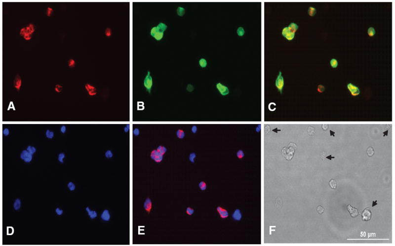
Fig. 1.

Dissociated CB cells were incubated with biotinylated PNA and labeled with Alexa 546 tagged streptoavidin (A) and double labeled by TH with Alexa 488 secondary antibody (B). PNA and TH are overlaid (C). The nucleus are stained with DAPI contained mounting medium (D) and PNA stained image is overlaid over DAPI (E). The CB cells labeled with PNA are exactly matched with cell labeled with TH. Thus PNA only labels type I cells and the cells marked with arrows on DIC (F) are not type I cells. A scale bar (50 μm) in DIC (F)
