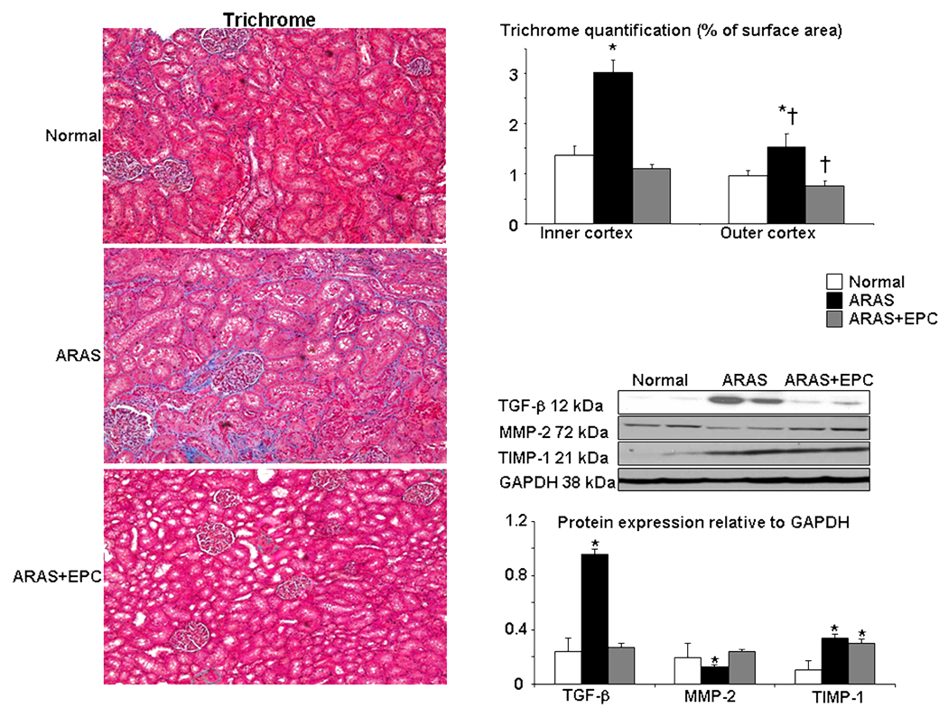
Figure 5.
Left: Representative renal trichrome staining (left, ×20) and quantification in the outer and inner renal cortex. Right: protein expression and quantification of transforming growth factor (TGF)-β, matrix-metalloproteinase (MMP)-2 and tissue-inhibitor of metalloproteinase (TIMP)-1 in normal, atherosclerotic renal artery stenosis (ARAS), and ARAS kidneys treated with endothelial progenitor cells (ARAS+EPC). EPC administration decreased renal TGF-β improved MMP-2, and attenuated renal fibrosis. *p<0.05 vs. Normal, † p<0.05 vs. Inner cortex

