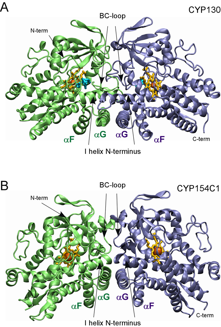
Fig. 5. Dimerization interface.
The dimerization interfaces of (A) CYP130 (2000 Å3) and (B) CYP154C1 (610 Å3) formed largely via interactions between the G helices in anti-parallel orientations, overlapping N-termini of the I helices, and multiple contacts in the BC-loop regions are shown. The monomers are colored in green and blue, heme is in yellow and econazole in (A) is in cyan.

