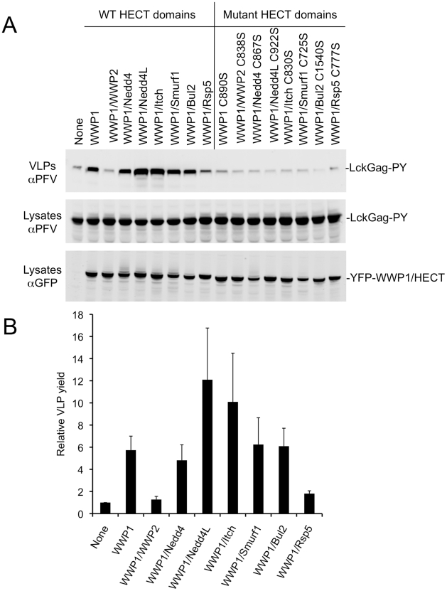
Figure 2. Stimulation of PPxY-dependent VLP production by chimeric HECT ubiquitin ligases.
(A) Quantitative Western blot (LICOR) analysis of VLP release from 293T cells co-expressing Lck-Gag-PY and either YFP alone (None) or the indicated YFP-fused WWP1 C2/WW domains linked to the indicated HECT domains. Note that the unfused YFP is not visible in the “None” lane because it migrates to a different position on the blot. (B) Quantitation of Lck-Gag-PY protein in particles by quantitative Western blot analysis (LICOR). Values plotted are the levels of VLP associated Lck-Gag-PY protein generated in the presence of the indicated YFP-fused chimeric ubiquitin ligase, relative to that generated in the presence of YFP only (None). Data represent the mean and standard deviation of four independent experiments.

