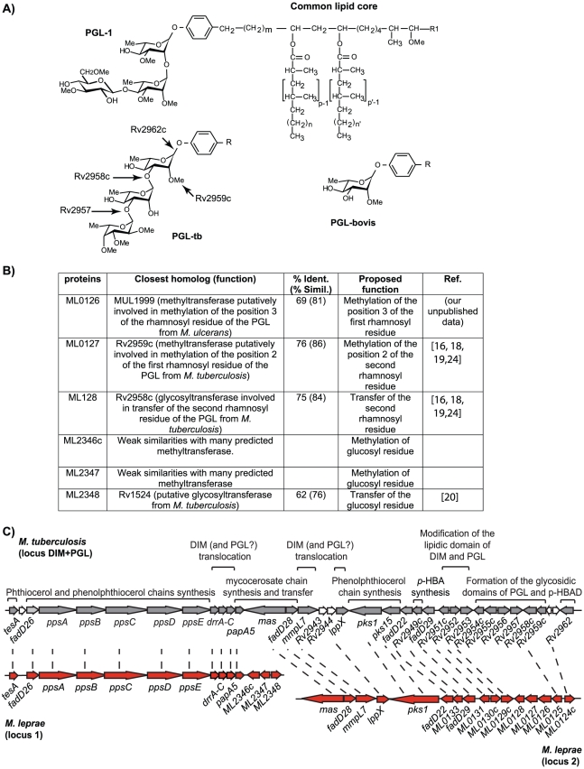
Figure 1. Identification of the genes involved in the formation of the saccharidic domain of PGL-1 from M. leprae.
(A) Structure of the PGL from M. leprae, M. tuberculosis and M. bovis and role of the various enzymes from M. tuberculosis in the formation of the saccharidic domain of PGL-tb. In M. leprae, p, p′ = 4; n, n′ = 16–20; m = 17 [5]; R1 = −CH2−CH3 or −CH3; R = common lipid core. (B) Candidate proteins for the formation of the terminal disaccharide of PGL-1 and proposed enzymatic function. (C) DIM+PGL loci in M. tuberculosis and in M. leprae. Orthologs are linked by dashed lines. Known functions of the encoded proteins are indicated above the open-reading frames.

