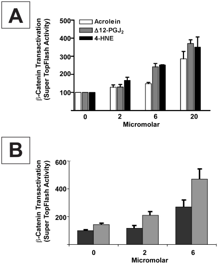
Figure 6. α, ß–unsaturated carbonyls enhance nuclear β-catenin signaling in STF3A cells:concentration-response relationships.
(A) Nuclear β-catenin signaling (luciferase luminescence) in STF3A cells (2×10⁴ cells/well) grown for 24 hrs in medium containing 0, 2, 6 and 20 µM each of acrolein (□), Δ12 PGJ2 (▪) and 4-HNE (▪). (B) Nuclear β-catenin signaling in STF3A cells (2×10⁴ cells/well) grown for 24 hrs in medium containing 0, 2 or 6 µM of Δ12 PGJ2 alone (▪) or Δ12 PGJ2 plus 100 µM BSO. GSH levels fell by 79% and 88% at 6 and 24 hrs after treating cells with BSO; 92–95% of cells were still viable at 24 hrs. Bars represent the mean ± s.e.m from n≥3 separate experiments.

