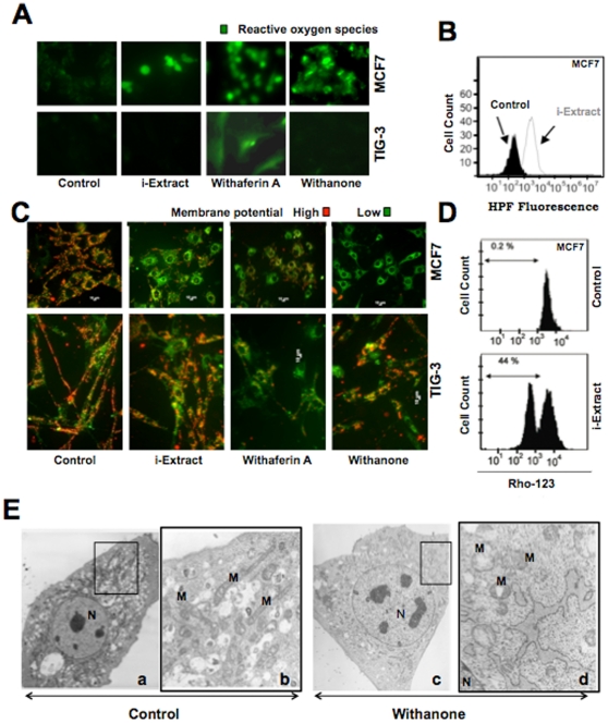
Figure 6. Role of ROS and mitochondrial damage in i-Extract induced cytotoxicity.
MCF7 cells showed the induction of ROS when treated with i-Extract, Withaferin A or Withanone (A and B). Normal cells showed ROS induction only in the presence of Withaferin A (A). Loss of mitochondrial membrane potential in MCF7 cancer cells, as seen by JC-1 staining was detected with i-Extract only (C). Preferential induction of loss of mitochondrial trans-membrane potential in MCF7 cells detected by flow cytometry using RHO-123 increased from 0.2% of population in control to 44% of population treated with i-Extract (D). Mitochondrial damage was detected in Withanone-treated MCF7 cells. (E), Transmission electron microscopic images of control and Withanone-treated MCF7 cells. Control cells showed normal elongated mitochondrial (M) with parallel cristae (a) (enlarged boxed image, b), Withanone-treated cells showing swollen mitochondria with reduced number of the cristae (c) (enlarged boxed image, d). N, Nucleus.

