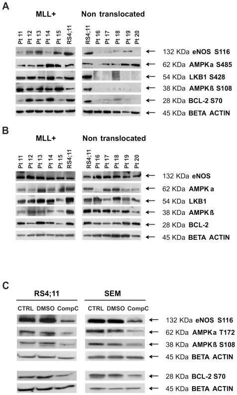
Figure 2. Validation of RPMA results through Western Blot.
(A) Hyperactivation of the AMPK pathway in MLL-rearranged patients vs non-translocated ones (independent sets of pediatric BCP-ALL at diagnosis: patients 11–15 are MLL-rearranged -all MLL-AF4-, patients 16–20 are non-translocated). RS4;11 cell lysate was used as positive control for antibody staining. (B) Total forms of the AMPK pathway proteins in previously described patients: 11–15 are MLL-rearranged and 16–20 are non-rearranged. RS4;11 cell lysate was used as positive control for antibody staining. There are no substantial differences on total protein form levels between MLL-rearranged and non-translocated patients. (C) AMPK pathway inhibition after Compound C treatment. RS4;11 and SEM cells (both MLL-rearranged) were treated with the AMPK inhibitor Compound C 8 µM for 48 hours. Phosphorylation of AMPK pathway proteins was evaluated through WB in control, DMSO treated and Compound C treated cells.

