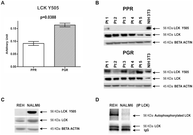
Figure 3. Hyperactivation of LCK in PPR patients.
(A) LCK Y505 measured with RPMA is higher in Prednisone Good Responder (PGR) (0.164±0.007) than in Prednisone Poor Responder (PPR) (0.092±0.008) patients (two-sample Welch t-statistics -unequal variances- with Bonferroni multiplicity corrections p = 0.0388). (B) LCK hyperphosphorylation at Y505 in PGR patients was confirmed by Western Blot in an independent set of specimens (pediatric BCP-ALL at diagnosis: patients 1–5 are Prednisone Good Responders, patients 6–10 are Prednisone Poor Responders). NIH3T3 commercial cell lysate (BD Biosciences) was used as positive control for antibody staining. Expression of the total form of LCK does not differ between the patients. (C) LCK activation state in the cell lines chosen as in vitro model. REH cell lines are a model for PPR patients, while NALM6 are a model for PPR patients. The LCK activation state was evaluated through WB. (D) In vitro LCK autophosphorylation assay. LCK was immunoprecipitated from REH and NALM6 cells, and then incubated in a phosphorylation mixture at 30°C for 40 min. Autophosphorylation of LCK was analyzed by digital autoradiography on Cyclone Plus (upper gel), and by WB with anti-LCK antibody to assess the LCK amount (lower gel).

