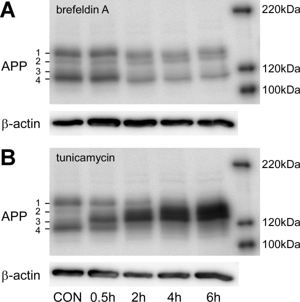
Figure 4.
Shifts in full length APP patterns induced by the glycosylation inhibitors tunicamycin and brefeldin A in mononuclear phagocyte cultures. Human mononuclear phagocytes were isolated as indicated and left unstimulated on ultra-low attachment plates for 3 days. 6 h, 4 h, 2 h or 0.5 h prior to the lysis of the cells, tunicamycin or brefeldin A were added in a concentration of 10 μg/ml each. Cells were lysed in RIPA buffer and APP expression was analysed by separation on 7.5% SDS-PAGE, subsequent blotting on PVDF-membranes and staining with the 1E8 monoclonal antibody. Staining of β-actin served as a loading control. The right hand side shows a molecular weight standard. Note the slight shift in molecular weight after brefeldin A treatment and the decrease of band APP1 corresponding to mature APP and the increased amounts of the bands APP2 and APP3 after treatment with Tunicamycin.

