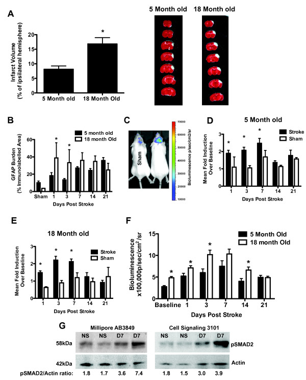
Figure 2.
TGFβ signaling in the brain increases with age and stroke. A.) Infarct volume and representative TTC images of the lesion 24 hours following dCMAO (*p < 0.05). B.) Quantification of GFAP immunostain, showing that astrogliosis is accelerated in aged mice (*p < 0.05 compared to 5 month old). C.) Representative bioluminescence showing reporter gene (luciferase) expression is increased after stroke compared to surgical shams (2 females shown at 3 days post stroke). D.) Time course of TGFβ signaling (luciferase induction) after stroke in 5 month old females (*p < 0.05 compared to sham). E.) Time course of TGFβ signaling (luciferase induction) after stroke in 18 month old females (*p < 0.05 compared to sham). F.) Comparison of absolute values of bioluminescence between young and old mice, showing that TGFβ signaling in the brain increases at baseline and after stroke (*p < 0.05 compared to 5 month old). G.) Western blot to corroborate that TGFβ signalling increases after stroke. Two different antibodies for pSmad2 were used to confirm that pSmad2 is increased at day 7 after stroke. NS = no stroke, D7 = day 7 post stroke. All error bars for this figure are SEM.

